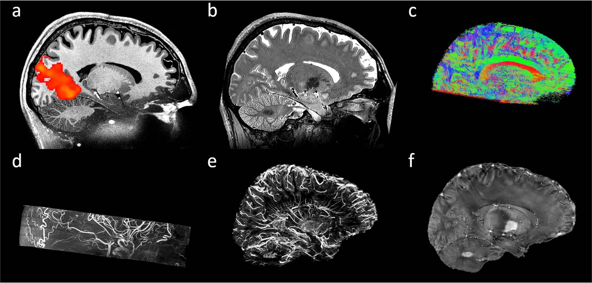

(b) An MRI machine generates a magnetic field around a patient. (a) The results of a CT scan of the head are shown as successive transverse sections. The term " noninvasive" is used to denote a procedure where no instrument is introduced into a patient's body, which is the case for most imaging techniques used. In the case of projectional radiography, the probe uses X-ray radiation, which is absorbed at different rates by different tissue types such as bone, muscle, and fat. In the case of medical ultrasound, the probe consists of ultrasonic pressure waves and echoes that go inside the tissue to show the internal structure. This means that cause (the properties of living tissue) is inferred from effect (the observed signal). In this restricted sense, medical imaging can be seen as the solution of mathematical inverse problems. Medical imaging is often perceived to designate the set of techniques that noninvasively produce images of the internal aspect of the body. As of 2015, annual shipments of medical imaging chips amount to 46 million units and $1.1 billion. Medical imaging equipment are manufactured using technology from the semiconductor industry, including CMOS integrated circuit chips, power semiconductor devices, sensors such as image sensors (particularly CMOS sensors) and biosensors, and processors such as microcontrollers, microprocessors, digital signal processors, media processors and system-on-chip devices. Radiation exposure from medical imaging in 2006 made up about 50% of total ionizing radiation exposure in the United States. In a limited comparison, these technologies can be considered forms of medical imaging in another discipline.Īs of 2010, 5 billion medical imaging studies had been conducted worldwide. time or maps that contain data about the measurement locations. Measurement and recording techniques that are not primarily designed to produce images, such as electroencephalography (EEG), magnetoencephalography (MEG), electrocardiography (ECG), and others, represent other technologies that produce data susceptible to representation as a parameter graph vs. Although imaging of removed organs and tissues can be performed for medical reasons, such procedures are usually considered part of pathology instead of medical imaging.Īs a discipline and in its widest sense, it is part of biological imaging and incorporates radiology, which uses the imaging technologies of X-ray radiography, magnetic resonance imaging, ultrasound, endoscopy, elastography, tactile imaging, thermography, medical photography, nuclear medicine functional imaging techniques as positron emission tomography (PET) and single-photon emission computed tomography (SPECT). Medical imaging also establishes a database of normal anatomy and physiology to make it possible to identify abnormalities.
#EFILM LITE EXPORT MRI DWI SKIN#
Medical imaging seeks to reveal internal structures hidden by the skin and bones, as well as to diagnose and treat disease. Medical imaging is the technique and process of imaging the interior of a body for clinical analysis and medical intervention, as well as visual representation of the function of some organs or tissues ( physiology).

Usually some value between 100% and 157% is chosen in clinical practice.A CT scan image showing a ruptured abdominal aortic aneurysm Scans with higher coverage factors have fewer artifacts and higher signal-to-noise, at the cost of increased imaging time. Scans using coverage factors lower than 100% have more noticeable streaking artifacts. A coverage factor of 157% would be gapless, while a coverage factor of 100% would not impose any time penalty compared to a Cartesian method but would contain gaps between the ends of the blades.

The degree of blade overlap (angle between the blades) is usually an operator-selectable parameter given as a percentage with a vendor-specific name such as "blade coverage factor" (Siemens), "MultiVane percentage" (Philips), or " k-space filling factor" (Toshiba). If there are L lines per blade and N blades, the equivalent imaging matrix diameter M is given by the equation LN = πM/2.Ĭomplete coverage of the k-space circle (without gaps between the blades) therefore requires a factor of π/2 ≈ 1.57 times as long as coverage using a Cartesian (rectangular) method. However, these locations only contribute high spatial frequency information at oblique angles within the final image, and so are often considered the "least important" regions of k-space. Although the center of k-space is highly oversampled, the "corners" of k-space are not sampled at all.


 0 kommentar(er)
